
Wet Macular Degeneration: Causes, Treatment, and What to Expect


What Is Wet Macular Degeneration
Wet macular degeneration is an advanced form of age-related macular degeneration (AMD), the leading cause of central vision loss in older adults. It develops when abnormal blood vessels grow beneath the retina and leak fluid or blood, damaging the macula, which is the small central portion of the retina you rely on for reading, driving, and recognizing faces.
Dry AMD is the more common form and develops gradually as tiny yellow deposits called drusen accumulate under the retina and the light-sensitive cells slowly break down over years. Wet AMD behaves very differently. It can cause severe central vision loss within weeks to months if left untreated. Any stage of dry AMD can convert to wet AMD, sometimes without noticeable warning, which is why regular monitoring matters so much.
The process that drives wet AMD is called choroidal neovascularization, meaning new blood vessels grow from a layer beneath the retina called the choroid. As the eye ages, reduced nutrient and oxygen flow prompts the retina to overproduce a protein called vascular endothelial growth factor (VEGF). High VEGF levels signal the body to grow new vessels, but these vessels are fragile and leak, disrupting the retina's delicate structure and causing rapid loss of the light-sensing cells called photoreceptors. Over time, leaking and scarring destroy central vision.

Who Is at Risk
Several factors increase the likelihood of developing wet AMD, including age, genetics, and certain lifestyle habits. Understanding your personal risk profile can help guide how often you should be screened and what steps to take.
Age is the strongest risk factor for wet AMD. The condition primarily affects people over 50, and the risk rises significantly with each decade. Approximately 200,000 new cases are diagnosed each year in North America, and having dry AMD in one or both eyes is itself a major risk factor for developing the wet form.
Genetics play a meaningful role in AMD. People with a first-degree relative who has AMD face roughly two and a half times the average risk of developing the condition. If dry AMD runs in your family, discussing your personal risk with an ophthalmologist is an important step toward early detection.
Several modifiable factors are associated with wet AMD, and addressing them may help slow disease progression. Smoking is among the most significant, increasing risk by two to three times.
- Cardiovascular disease and high blood pressure raise AMD risk by approximately one and a half times
- High cholesterol is also associated with increased risk
- Obesity may double the risk of AMD progression
- Prolonged ultraviolet light exposure without protective eyewear is also a contributing factor
People of Caucasian descent have a higher prevalence of wet AMD compared to other racial groups. Women are affected more frequently than men. People with lighter-colored eyes may also face slightly increased risk due to lower natural pigment protection in the retina.

Signs and Symptoms
Wet AMD can develop suddenly, and its symptoms are distinct from other common eye conditions. Recognizing the warning signs early and acting quickly is critical, because damage that occurs before treatment begins is often permanent.
The most common early symptom is the appearance of straight lines looking wavy or bent, a distortion called metamorphopsia. This is a hallmark sign of wet AMD and should prompt immediate evaluation. Other early signs include a dark or blank spot in the center of your vision, difficulty recognizing faces, trouble reading even with corrective lenses, and colors appearing less vivid than usual.
Certain vision changes should be treated as an eye emergency. Contact a retina specialist or seek urgent care immediately if you notice any of the following.
- Sudden blurring or a rapid decrease in central vision
- Straight lines appearing wavy, bent, or distorted
- A new dark or blank area in your central vision
- Any abrupt change in the quality of your vision
Because wet AMD can worsen within days, delaying evaluation even briefly can lead to additional irreversible vision loss. Patients already diagnosed with dry AMD should check their vision daily using an Amsler grid, a simple chart of straight lines with a dot at the center that helps detect subtle distortion early.
Wet AMD often affects one eye before the other. When only one eye is involved, the unaffected eye often compensates, making it difficult to notice the change. This is why checking each eye separately while using an Amsler grid is so important. Covering one eye at a time can reveal distortion that would otherwise go undetected.
Diagnosis and Testing
Accurately diagnosing wet AMD requires a combination of a thorough eye examination and specialized imaging. These tests help confirm the diagnosis, locate abnormal blood vessels, and guide treatment decisions.
Diagnosis begins with a comprehensive dilated eye exam. Drops are placed in the eye to widen the pupil, allowing a clear view of the retina and macula. During this exam, the specialist looks for signs of abnormal blood vessels, fluid, bleeding, or scarring beneath the retina.
Optical coherence tomography (OCT) is one of the most important tools for diagnosing and monitoring wet AMD. It uses light waves to create detailed cross-sectional images of the retina, revealing fluid under or within the retinal layers, swelling of the macula, and changes in retinal thickness. The test takes only a few minutes and is routinely used to track how well treatment is working.
Fluorescein angiography maps the location and extent of abnormal blood vessels. A special dye is injected into a vein in the arm and circulates to the blood vessels of the eye, where a camera captures images showing where leakage is occurring. This information helps the retina specialist determine the best course of treatment.
OCT angiography (OCTA) is a newer imaging technique that visualizes blood flow in the retina without the need for a dye injection. It provides detailed maps of retinal and choroidal blood vessels and is particularly useful for ongoing monitoring and for detecting abnormal vessel growth in its earliest stages.

Treatment Options for Wet AMD
The goal of treatment is to stop the leaking of abnormal blood vessels, reduce damage to the macula, and preserve as much central vision as possible. Several well-established and newer options are available, and treatment is tailored to each patient's needs and response.
Anti-VEGF injections are the standard treatment for wet AMD. These medications block VEGF, the protein that drives abnormal blood vessel growth and leakage. They are delivered as intravitreal injections, meaning the medication is injected directly into the vitreous, the gel-like fluid inside the eye. Treatment typically begins with monthly injections during an initial loading phase, after which the interval may be extended based on how the eye responds. With consistent treatment, anti-VEGF therapy can stabilize vision in the large majority of patients and improve vision in a meaningful portion of them.
Several anti-VEGF medications are approved for treating wet AMD, and each has its own dosing schedule and characteristics.
- Lucentis (ranibizumab): One of the first anti-VEGF treatments approved for wet AMD, typically given every four weeks
- Eylea (aflibercept): Commonly used, with dosing that can often be extended to every four to eight weeks after initial treatment
- Eylea HD (high-dose aflibercept): A higher-concentration formulation that may allow dosing intervals of eight weeks or longer
- Vabysmo (faricimab): A bispecific antibody targeting both VEGF and another pathway called angiopoietin-2, which allows many patients to extend dosing to every 12 to 16 weeks
- Beovu (brolucizumab): Dosed every eight to twelve weeks, though it carries a risk of retinal vasculitis, which is inflammation of the retinal blood vessels
- Avastin (bevacizumab): Used off-label in retina care due to its effectiveness, though it is FDA-approved for cancer treatment rather than eye disease
The Susvimo port delivery system is a surgically placed implant in the wall of the eye that continuously releases ranibizumab into the vitreous over several months. Rather than returning monthly for injections, patients typically need refills only about twice per year. This option may be well-suited for patients who respond well to ranibizumab but prefer fewer office visits. A retina specialist can determine if this approach is appropriate.
While anti-VEGF injections are the primary treatment, other approaches may be used in specific situations. Photodynamic therapy (PDT) uses a light-activated drug injected into the bloodstream that is then activated by a special laser aimed at the abnormal vessels. Laser photocoagulation uses thermal laser energy to seal leaking vessels. These approaches are less commonly used today but may be considered in certain cases or used alongside anti-VEGF therapy.

What Treatment Involves
Understanding what to expect during and after treatment helps many patients feel more prepared and confident about managing wet AMD long term.
Intravitreal injections are performed in the office. The eye is numbed with anesthetic drops or a small injection of numbing medicine, and the surface of the eye is cleaned with an antiseptic solution to reduce the risk of infection. The injection itself takes only a few seconds. Most patients feel pressure during the procedure and possibly mild discomfort. Temporary blurring, mild redness, and a feeling of grittiness are common afterward and typically resolve within a day or two.
Wet AMD requires ongoing treatment and monitoring over time. After the initial loading phase, the retina specialist uses OCT imaging and clinical exams to evaluate the eye's response and determine whether injection intervals can be safely extended. Some patients may eventually go several months between treatments, while others need more frequent visits to keep the disease under control. Missing or delaying scheduled appointments can allow fluid to return and cause additional vision loss.
Wet AMD is a chronic condition that requires long-term management. With consistent treatment, many patients are able to maintain useful central vision for years. The degree of improvement depends significantly on how much damage occurred before treatment began, which is why early diagnosis and prompt care offer the best possible outcome. Vision lost to scarring before treatment starts generally cannot be recovered.
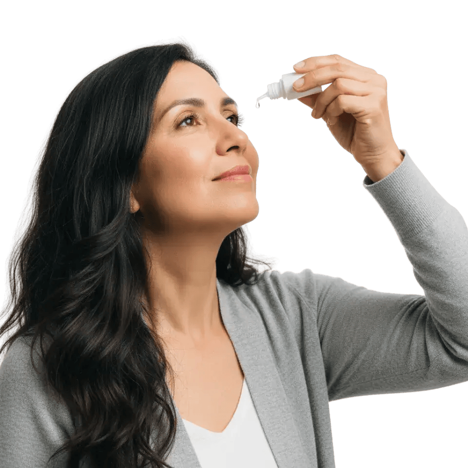
Living with Wet AMD
Managing wet AMD goes beyond office visits. Daily habits, self-monitoring tools, and lifestyle choices all play a role in preserving your vision and maintaining quality of life.
An Amsler grid is a simple chart with a grid of straight lines and a dot at the center. Checking it daily, one eye at a time, helps detect new distortion, wavy lines, or blank spots that may signal disease activity. Any new changes should be reported to a retina specialist promptly, as early retreatment helps protect vision.
Even with treatment, some patients experience lasting changes to central vision. Low vision specialists can recommend tools and strategies that help maintain independence. Magnifying glasses, large-print materials, high-contrast device settings, audiobooks, task lighting, and glare reduction in the home are all commonly used resources that can make daily life more manageable.
Certain lifestyle changes may help slow AMD progression. Quitting smoking is one of the most impactful steps. A diet rich in leafy green vegetables, fish, and foods high in omega-3 fatty acids may support eye health. Regular exercise, maintaining a healthy weight, managing cardiovascular health, and wearing sunglasses with ultraviolet protection are also recommended. A specific combination of vitamins and minerals known as AREDS2 supplements has been shown to reduce the risk of advanced AMD progression in certain patients. A retina specialist can advise whether these supplements are appropriate based on the stage and type of AMD present.

When and Why to See a Specialist
Wet AMD requires specialized care from a retina specialist, also known as a vitreoretinal surgeon, who has advanced training in diagnosing and treating diseases of the retina. Knowing when to seek that care makes a significant difference.
Optometrists and general ophthalmologists often detect early signs of AMD during routine eye exams. If wet AMD is suspected, patients are referred to a retina specialist for further evaluation and treatment. Getting that referral promptly rather than waiting to see if symptoms worsen is always the right decision with this condition.
Patients diagnosed with wet AMD need consistent follow-up visits that include imaging to check for fluid or new vessel growth. Wet AMD can reactivate at any time, even when vision feels stable, so keeping all scheduled appointments is essential. Any sudden change in vision between appointments should be reported to a retina specialist right away rather than waiting for the next scheduled visit.

Frequently Asked Questions
Here are answers to some of the questions patients ask most often about wet AMD, including guidance on what to do and what to expect.
While there is currently no cure for wet AMD, anti-VEGF treatment can control the condition effectively in many patients. The key is consistency. Stopping treatment without medical guidance often leads to disease reactivation and further vision loss. Your retina specialist will work with you to find the safest dosing interval that keeps the condition stable over time.
Wet AMD affects central vision but does not cause total blindness, because peripheral (side) vision is preserved. Even in advanced cases, most patients retain enough side vision to move around safely and remain oriented. Modern anti-VEGF treatment has significantly reduced the risk of severe central vision loss compared to earlier decades, especially when treatment begins promptly.
The most reliable early indicator is a change detected on your daily Amsler grid check, particularly new waviness, distortion, or a blank spot. Because this conversion can happen without pain or dramatic symptoms, relying on daily self-monitoring and keeping scheduled appointments is critical. If something changes between visits, contact your retina specialist the same day rather than waiting.
Yes, and this is an active area of development. Vabysmo (faricimab) targets two disease pathways and has enabled many patients to extend treatment to every 12 to 16 weeks. Eylea HD also supports extended dosing intervals. The Susvimo port delivery system provides continuous drug release with refills approximately twice per year. Researchers are also investigating gene therapy approaches that may eventually reduce treatment burden further. Your retina specialist can discuss which of these options may be appropriate for your situation.
AREDS2 supplements are not a prevention tool for people with healthy eyes or early AMD. They are specifically intended for patients with intermediate AMD or advanced AMD in one eye, where the evidence shows they can reduce the risk of progression to advanced AMD. They are not appropriate for everyone, and taking them without a recommendation from a retina specialist is not advised. If you have been diagnosed with AMD, ask your specialist whether your stage qualifies.
Sudden changes in central vision, new distortion on the Amsler grid, or a new blank spot should be treated as an urgent situation. Contact a retina specialist the same day for evaluation. If you cannot reach your specialist, go to an emergency eye care facility. Even a delay of a day or two can allow significant additional damage when wet AMD is actively leaking, so prompt action is always the right choice.
Expert Wet AMD Care in New Jersey
At EyeCare Consultants of NJ, our team of ophthalmologists and retina specialists is dedicated to providing exceptional, personalized care for patients with wet macular degeneration. We combine advanced diagnostic imaging with the latest treatment options to help protect your central vision and your quality of life. We welcome patients from across New Jersey and encourage you to schedule an evaluation if you have any concerns about your vision or your risk for AMD.

Dr. Arnett has provided excellent medical attention for my retina eye surgery and has a very professional and positive bedside manner. I would highly recommend him.
R. T
Highly recommend Dr. Kayserman. Extremely competent, experienced, highly personable
LS
Dr. Larisa Kayzerman is a wonderful doctor. She is very attentive. Doctor maid operation the same day. I'm very satisfied with the result.
Rafail
Dr. Kayserman was gentle, efficient and provided extremely valuable information which was comforting to me. I felt better after seeing her and am looking forward to additional visits.
Lina N.
Dr. Arnett deserves 10 stars. He is the best.
AJ